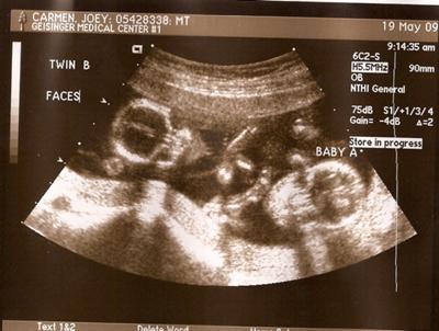
An ultrasound take a look at additionally referred to as “sonography” is a diagnostic medical imaging method, which is carried out to view a patient’s inside organs and to evaluate his or her blood stream by way of varied vessels. Moms to be may decide that getting a 3D ultrasound is a budget alternative to a more substantial prenatal care program and should discontinue this prenatal care under the assumption “all the pieces is ok.” While an excellent tool for determining the bodily situation of an unborn baby.
It is possible to be able to have a child gender ultrasound earlier than 20 weeks, around 15 weeks is usually ok. If you really want to know then the particular person performing the baby gender ultrasound should be capable of tell you, though it’s possible you’ll find it difficult to determine for your self.
In case a lady is pregnant, she may be asked by the physician to endure Doppler Sonography for a variety of reasons like measuring the movement of blood by way of the umbilical twine along with a correct analysis of the mind and coronary heart of the developing fetus.
Because of the ever rising demand for safer environments and procedures, sonography should proceed to develop. Different situations that prohibit using ultrasound remedy embrace the spine space of patients who’ve had spinal surgeries corresponding to Laminectomy, anyplace on a affected person the place anesthetics are being used, or any form of disposition to bleeding excessively.
Within the case of the widespread and potentially, major problem of blood clots in the deep veins of the leg, ultrasound plays a key diagnostic position, while ultrasonography of continual venous insufficiency of the legs focuses on more superficial veins to help with planning of appropriate interventions to alleviate symptoms or improve cosmetics.
Transcranial Doppler (TCD) and transcranial coloration Doppler (TCCD), which measure the velocity of blood movement via the mind ‘s blood vessels transcranially (by the skull ). They are used as checks to help diagnose emboli , stenosis , vasospasm from a subarachnoid hemorrhage (bleeding from a ruptured aneurysm ), and other problems.
In preparation for examination of the child and womb during being pregnant, typically it is suggested that moms drink at least four to six glasses of water approximately one to two hours previous to the examination for the aim of filling the bladder.
Diagnostic ultrasound, additionally referred to as sonography or diagnostic medical sonography, is an imaging technique that uses high-frequency sound waves to produce pictures of structures inside your physique. Léandre Pourcelot, who was a researcher and trainer at INSA (Institut Nationwide des Sciences Appliquées) Lyon copublished in 1965 a report at “Académie des sciences” « Effet Doppler et mesure du débit sanguin » (Doppler effect and measure of the blood circulation), the basis of his design of a Doppler move meter in 1967.
A radiologist, a doctor particularly trained to supervise and interpret radiology examinations, will analyze the pictures and send a signed report to your major care doctor, or to the doctor or different healthcare supplier who requested the examination.
Three-dimensional imaging permits a physician to get a greater look at the organ being examined and is finest used for early detection of tumors, discovering plenty within the colon and rectum, detecting breast lesions for possible biopsies, assessing the event of a fetus, and visualizing blood flow in numerous organs.
Medical ultrasound (also called diagnostic sonography or ultrasonography) is a diagnostic imaging approach based on the applying of ultrasound It’s used to create an image of inside body buildings corresponding to tendons , muscles , joints, blood vessels, and internal organs.
Ultrasound
At 16 to 20 weeks, a pregnant woman has the opportunity to search out out the gender of her child with the first ultrasound visit. You can be mendacity down with a full bladder for the process then high frequency waves cross via your body abdominal area with a medical system move backwards and forwards in the abdomen with a skinny jellylike substance applying to the skin in your stomach to enhance the contact of your body.
Medical doctors employ ultrasound imaging in diagnosing a wide variety of circumstances affecting the organs and soft tissues of the body, including the heart and blood vessels, liver , gallbladder , spleen , pancreas , kidneys , bladder , uterus, ovaries, eyes , thyroid , and testicles.
How Ultrasound Works
The physician spreads a lubricating gel on the realm being examined after which presses the transducer towards the skin to obtain photos.
With a population of more than three million individuals, Orange County, California is a pleasant mix of seashores, parks and properly-identified cities like Santa Ana, Newport Beach and Anaheim. One of the most frequent makes use of of ultrasound is during being pregnant, to monitor the expansion and improvement of the fetus, however there are various different uses, including imaging the guts, blood vessels, eyes, thyroid, mind, breast, stomach organs, pores and skin, and muscle tissue.
There are a number of which can be more hardy or cellular and may be more difficult to treat, but when ultrasound is mixed with something like pond micro organism, which is designed to reduce nutrients within the water, most algae will have a troublesome time in surviving and rising.
Superficial buildings such as muscle , tendon , testis , breast , thyroid and parathyroid glands, and the neonatal mind are imaged at a better frequency (7-18 MHz), which provides better linear (axial) and horizontal (lateral) resolution Deeper buildings comparable to liver and kidney are imaged at a decrease frequency 1-6 MHz with lower axial and lateral decision as a price of deeper tissue penetration.
In the event you look closely at the The American Registry for Diagnostic Medical Sonography (ARDMS) prerequisites for getting licensed as an ultrasound technologist, you’ll notice that nowhere does it state that it’s good to go to a licensed ultrasound technologist program like those offered by Michener and Mohawk.
Sonographers
Medical ultrasound falls into two distinct categories: diagnostic and therapeutic. Ultrasound is sound waves with frequencies higher than the higher audible limit of human hearing Ultrasound shouldn’t be totally different from “regular” (audible) sound in its bodily properties, besides that humans cannot hear it. This restrict varies from person to person and is approximately 20 kilohertz (20,000 hertz) in healthy younger adults.
Should you select to go into normal radiography you’ll be able to opt to enter into diagnostic sonography or nuclear medication later on. Basic sonographers use x-ray, magnetic resonance imaging, computed tomography and mammography applied sciences to take footage of the within of sufferers’ bodies to diagnose illness, deal with medical conditions and look at fetuses in pregnant girls.
Function, Process, And Preparation
If you want to discover ways to get into a sonography faculty, first you must determine what sort of sonography career you need to have. An ultrasound scan makes use of high-frequency sound waves to make an image of a person’s inner physique constructions. Ultrasound could also be used to assist physicians guide needles into the physique. Ultrasound transducers will be used in keeping with the research and how deep the tissue is that is being visualized.
Generally, doctors will use ultrasound scanning to watch and information invasive procedures like a biopsy of an individual’s breast or thyroid gland. In a transvaginal ultrasound , a transducer wand is placed in a woman’s vagina to get higher pictures of her uterus and ovaries.
sop ultrasound terapi, ultrasound adalah pdf, ultrasound atomization humidifier
A Sonographer can also be known as an Ultrasound Technician or a Diagnostic Medical Sonographer. HIFU is being investigated as a way for modifying or destroying diseased or abnormal tissues inside the physique (e.g. tumors) without having to open or tear the pores and skin or trigger harm to the encircling tissue. 3D ultrasound scans are as secure as traditional ultrasound scanning as a result of the picture is composed of sections of two-dimensional photographs, so the ultrasound publicity is similar.

